
Our Services
MRI - Magnetic Resonance Imaging
MRI uses a strong magnetic field and low-energy radiofrequency waves to examine a specific area of your body.
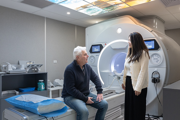

What is MRI - Magnetic Resonance Imaging?
Imaging @ Olympic Park has three Medicare licensed 3T 70cm bore MRI scanners. MRI uses a strong magnetic field and low-energy radiofrequency waves to examine a specific area of your body. The frequency of the radiofrequency waves used in MRI is similar to those used for FM radio stations.
The main advantage of MRI compared with other imaging modalities is that it provides the highest soft tissue resolution. Therefore, body structures such as the brain, nerves, tendons, ligaments, cartilage, spinal cord, breast, and the abdomen are highly suited to MRI examinations.
MRI is painless and does not use ionising radiation.
What to expect from an MRI appointment
Our MRI technologist will escort you to the scanning suite and prepare you for your scan. Preparation may include getting changed into a gown and removing items such as phones, watches and jewellery prior to entering the scan room.
The MRI technologist will ensure you are as comfortable as possible prior to starting the examination as you will need to remain still for the entire scan. If you are unsettled, it is important you communicate this to our staff, so they can adjust your position or use additional soft padding to ensure your examination is as pleasant and relaxing as possible. Most MRI scans take 10-15 minutes per body part.
All three of our MRI machines have a 70cm wide bore with lighting and a fan for ventilation. Some patients may experience claustrophobia, and our MRI technologists are very experienced at managing these situations. Just be sure to let us know if you are claustrophobic at time of booking and on your paperwork prior to your scan and we’ll ensure to make it is as comfortable as we can for you.
The MRI will make loud, rhythmic, buzzing and banging noises throughout your examination. Generally, patients do not find this unpleasant. Ear plugs will be given to you for hearing protection and you will also be given headphones over the top to listen to music during your scan. The MRI technologist will also talk to you through your headphones during the scan and you will be able to talk back to them. You may be given an emergency buzzer which can be used to stop the examination in case you need to communicate with the MRI technologist urgently, and if necessary, they can remove you from the scanner quickly.
Sometimes, an intravenous contrast injection of gadolinium may be required to make your examination more detailed. Gadolinium is a paramagnetic material which highlights various structures and pathologies in the MRI image to provide more detailed information.
How to prepare for your appointment
When making your appointment, you will be asked a series of safety questions to ensure you are safe to be scanned. You will then complete a more comprehensive safety questionnaire on arrival to your appointment.
Wear comfortable clothing to your appointment.
Our team will confirm any other specific requirements prior to your appointment, but generally no preparation is necessary.
What do you need to bring to your appointment?
- Your referral form – please note we accept all referral forms from all providers.
- Medicare card
- Pension or concession card
- Any prior imaging
When will my doctor receive the results?
On completion of the scan, our Radiologist will examine the images and send a report directly to your doctor. This usually takes between 24 hours to a week. You will then follow up with your referrer.
Your images and report will also be available to you via our patient portal.